Background and Significance
In life sciences research, cells and their interactions form the foundation of complex life activities. From the migration and coordination of immune cells to the encoding and transmission of information within neural networks, these processes typically occur within intact physiological environments and evolve continuously over extended periods. Compared to in vitro experimental conditions, cellular behaviors in living organisms are more authentic and highly dynamic. Therefore, achieving high-resolution, long-term, stable, and reliable 3D imaging under intravital conditions is of paramount importance for understanding the essence of biological processes.
However, this objective has long been severely hindered by optical aberrations. In living tissues or complex biological environments, inhomogeneous refractive index distributions cause wavefront distortions as fluorescent signals propagate, leading to blurred images, distorted structures, and even a loss of information. Aberrations not only degrade image quality but also introduce systematic errors into subsequent quantitative analyses, such as cell trajectory extraction, neural signal analysis, and population behavior modeling. Consequently, achieving high-precision aberration correction in complex intravital environments has become a critical scientific challenge driving the development of microscopic imaging technology.
Challenges in Existing Methods
To address this issue, adaptive optics (AO) methods have been widely introduced into biological imaging.
·Traditional Hardware AO: Directly modulates the wavefront using optical components like deformable mirrors; however, the systems are complex, costly, and limited in adjustment speed.
·Computational AO: Attempts to infer aberrations from image data and perform digital corrections. While offering greater flexibility, these methods often lack sufficient precision when dealing with large aberrations or complex samples in practical applications.
·Information Incompleteness: Existing methods generally fail to fully utilize multi-angle information, leaving wavefront estimation under-constrained.
In recent years, the development of light-field microscopy has offered new insights. Light-field microscopy simultaneously captures spatial and angular information, providing potentially rich dimensions for aberration estimation. Nevertheless, effectively extracting wave-optics information from high-dimensional spatial-angular data and applying it to high-precision aberration recovery remains an unresolved challenge.
The LEAO Method
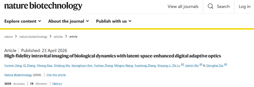
Addressing these challenges, the collaborative research team proposed a latent-space-enhanced digital adaptive optics (LEAO) method, published in Nature Biotechnology (Impact Factor: 41.7). LEAO introduces a new paradigm that integrates physical modeling with deep learning. By leveraging the semantic representation heterogeneity between the aberration wavefront and sample structure within a latent space, it achieves precise extraction and decoupling of the two. This enables researchers to observe biological processes under conditions much closer to their true physiological states. The method holds broad application prospects across multiple fields, including neuroscience and immunology, and is poised to propel microscopic imaging into a new era of high-fidelity observation.
Algorithmic Implementation
In terms of algorithm design, LEAO preserves the continuous wave properties of light-field data, treating it as a high-dimensional observation that encodes complete wavefront information.
1.Latent Space Mapping: LEAO first maps the original spatial-angular measurements into a high-dimensional latent space using an autoencoder network. Unlike direct regression, this latent space is designed with explicit physical semantics: one set of dimensions primarily encodes sample structural information, while the other encodes the aberration wavefront.
2.Triplet-Based Training: To achieve latent space decoupling and contrastive feature learning, the model introduces a triplet-based training strategy. A triplet consists of data where two samples share the same structure but have different aberrations, while two samples share the same aberration but have different structures. For samples with identical aberrations/structures, their corresponding aberration/structural feature vectors are pulled closer in the latent space. Conversely, for samples with different aberrations/structures, their feature vectors are pushed apart. This strategy allows physical information representations to form stable manifold structures within the latent space, thereby minimizing the interference of structural variations on aberration estimation.
3.Wavefront Phase Distribution: Once the latent space representation is obtained, a custom-designed estimator network extracts aberration-related features and outputs a continuous wavefront phase distribution. This wavefront is then used to generate a point spread function (PSF) that conforms to the wave-optics propagation model.
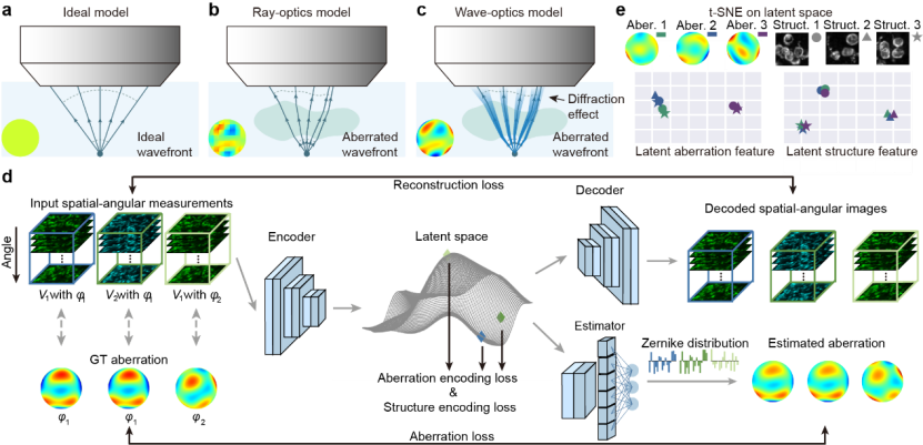
(Figure 1: Principle of LEAO)
Key Advantage: Unlike traditional methods that require repetitive PSF calculations, LEAO achieves stable wavefront estimation via a single forward inference pass, significantly improving computational efficiency.
Performance and Applications
1. Superior Performance and Robustness
LEAO delivers performance that is significantly superior to existing methods. Under large aberration conditions (ranging from 1 to 5 wavelengths), LEAO's aberration estimation accuracy is markedly improved compared to existing methods, maintaining stable performance in complex scenarios. Furthermore, under extremely low signal-to-noise ratio conditions (as low as 3.4 dB), LEAO maintains high-precision estimation where traditional methods fail noticeably. This demonstrates LEAO’s enhanced robustness under photon-limited conditions. Additionally, LEAO exhibits excellent adaptability to variations in angular sampling numbers, spatial sampling rates, and different imaging systems, working stably across diverse light-field microscopy configurations.
2. Cortical Imaging & Neural Network Research
Simultaneous imaging across multiple regions of the mouse cerebral cortex introduces spatially non-uniform, complex aberrations that are difficult for traditional methods to correct. Employing a "divide-and-conquer" approach, LEAO precisely identifies spatially varying aberrations across different regions. This helped researchers successfully restore the true morphology and distribution of neurons, significantly increasing the number of identifiable neurons and yielding more reliable functional signals. This work provides a powerful new technological foundation for mesoscale neural network functional research.
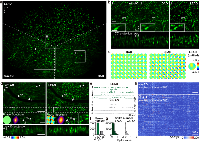
(Figure 2: LEAO corrects spatially non-uniform aberrations to enhance the contrast of neuronal calcium signals)
3. Intact-Skull Brain Imaging & Immune Microenvironment
In even more challenging intact-skull brain imaging experiments, LEAO enabled long-term monitoring of neutrophil behavior in mice subjected to traumatic brain injury (TBI) models. To better study the post-injury immune microenvironment, the curved skull structure was kept intact, which introduced massive optical aberrations into the imaging process.
Using LEAO, researchers recorded—for the first time—the entire process of activation, extravasation, and migration of thousands of neutrophils. These dynamic processes were previously impossible to observe directly due to severe aberrations. LEAO not only restored the image structures but also enabled accurate quantification of cell density and kinetic parameters.
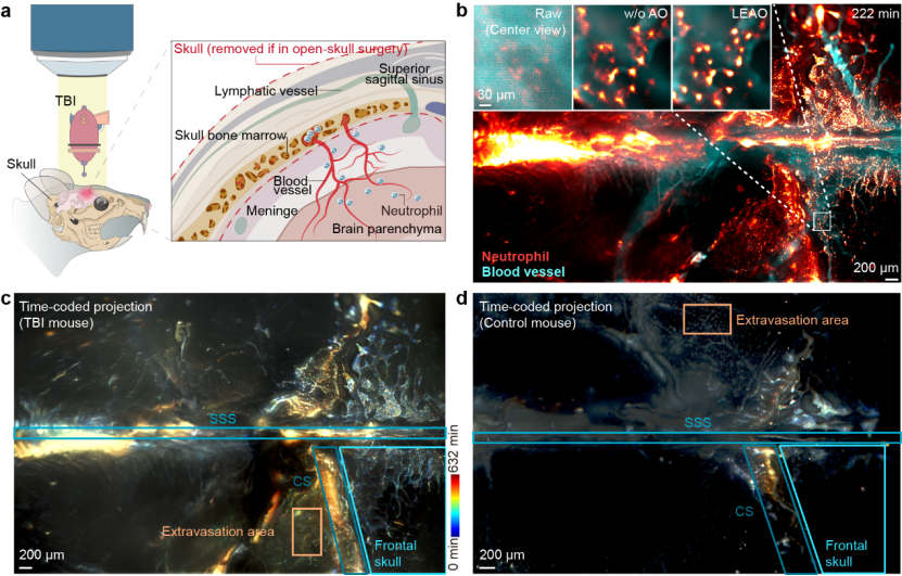
(Figure 3: Comparison of neutrophil activity between TBI mice and control mice)
Authorship and Support
·Co-Corresponding Authors: Academician Qionghai Dai (Department of Automation, Tsinghua University), Associate Professor Jiamin Wu (Department of Automation, Tsinghua University), and Assistant Professor Zhi Lu (Department of Psychology and Cognitive Sciences, Tsinghua University).
·First Author: Yunmin Zeng, a PhD student from the Department of Automation.
·Contributors: Qi Zhang, Yihong Xiao, Shidong Wu, Seonghoon Kim, Yunhao Zhang, Mingrui Wang, Yuanlong Zhang, and Xinyang Li made vital contributions to this work.
Funding and Support: This work was strongly supported by the National Natural Science Foundation of China, the Beijing Natural Science Foundation, the National Postdoctoral Program for Innovative Talents, the China Postdoctoral Science Foundation, the Tsinghua Shuimu Scholar Program, and the Tsinghua-Peking Center for Life Sciences.
Paper Link: https://www.nature.com/articles/s41587-026-03107-2
Faculty Profile:
Zhi Lu

Position: Assistant Professor, Doctoral Advisor, Department of Psychological and Cognitive Sciences
Research Area: Mesoscale Intravital Microscopic Imaging and Artificial Intelligence